Published July 2021
Jennifer Doran, MD 1, Josephine Cool 2
1 Resident, Internal Medicine, University of Colorado, 2 Instructor at Harvard Medical School, Director of Procedural and Ultrasound Education in Hospital Medicine and Director of Ultrasound Education for Internal Medicine Residency, Beth Israel Deaconess Medical Center
Objective(s)
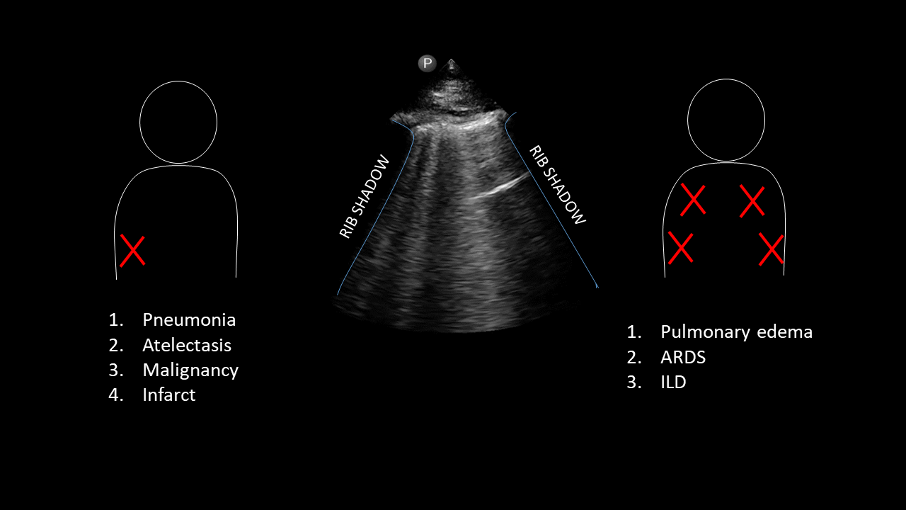
- Identify B-lines on lung ultrasound and describe the criteria for “B-profile” in a given window.
- Generate a differential based on clinical history and distribution of B-lines.
Teaching Instructions
Plan to spend 5-10 minutes familiarizing yourself with the animations of the PowerPoint and the key findings of this POCUS clip.
Instructions: Download the PowerPoint presentation file (videos do not work in the browser viewer) and have the image pulled up in presenter mode before learners look at the screen, to avoid revealing the diagnosis. Ask learners to discuss with their neighbors the answer to each question for about 30 seconds. Then advance through the animations and teaching points to the next question, and repeat. You can go back to prior graphics and questions by using the back arrow on the keyboard or by scrolling back on the mouse wheel. This should take no more than 5-10 mintues.
Diagnosis: Likely congestive heart failure exacerbation.
Teaching:
B-lines represent any increased density (scarring, water, pus, blood, or protein) in the alveoli or interstitium immediately deep to the visceral pleura. Instead of being reflected/deflected from aerated lung, the ultrasound waves penetrate the denser tissue, reverberate in that space then return to the transducer. This produces a comet-like artifact from the pleura to the bottom of the screen that moves with respirations.
Presentation Board
Take Home Point
- B-lines are reverberation artifacts that represent increased density in the lung alveoli or interstitium and appear as vertical comet-like beams from the pleura to the bottom of the screen and move with respirations.
- B-profile is consistent with cardiogenic edema in a patient presenting with left heart failure symptoms but could also represent other diffuse lung diseases.
References
Soni NJ. Point of care ultrasound. 2nd edition. ed. St. Louis, MO: Elsevier; 2019