Chalk Talks
Cardiology
Endocrinology
Gastroenterology
Hematology
Immunology
Infectious Disease
Nephrology
Neurology
Oncology
Primary Care
Pulmonology
Rheumatology
Transplant Medicine
Women’s Health
Procedures
Curricula Overviews
Procedures
POCUS
5-minute Cases
Case Conferences
Workshops
CXR
Case Conferences
Cavitary Lesion
Interstitial Lung Disease
Mediastinum and Hila
Obstructive lung diseases
Opacities
Pleural
Tubes, Lines, and Drains
ECG
Atrioventricular (AV) Heart Blocks
Bradycardias
Bundle Branch Block
Case Conferences
Electrolyte Abnormalities
Introduction
ST changes
Supraventricular Tachycardia
Dermatology
About
About Us
Contact
Twitter
Chalk Talks
Cardiology
Endocrinology
Gastroenterology
Hematology
Immunology
Infectious Disease
Nephrology
Neurology
Oncology
Primary Care
Pulmonology
Rheumatology
Transplant Medicine
Women’s Health
Chest x-ray
Case Conferences
Cavitary Lesion
Interstitial Lung Disease
Mediastinum and Hila
Obstructive lung diseases
Opacities
Pleural
Tubes, Lines, and Drains
ECGs
Atrioventricular (AV) Heart Blocks
Bradycardias
Bundle Branch Block
Case Conferences
Electrolyte Abnormalities
Introduction
ST changes
Supraventricular Tachycardia
POCUS
5-minute Cases
Case Conferences
Workshops
Procedures
Curricula Overviews
Procedures
About Us
Contact
POCUS
5-minute Cases
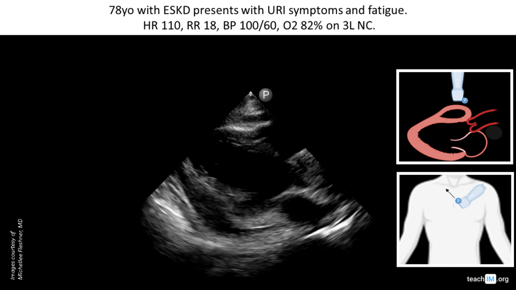
Pericardial Effusion Without Tamponade – POCUS
Identify a pericardial effusion in the parasternal long (PLAX) and assess for clinical signs of tamponade. (5 minutes)
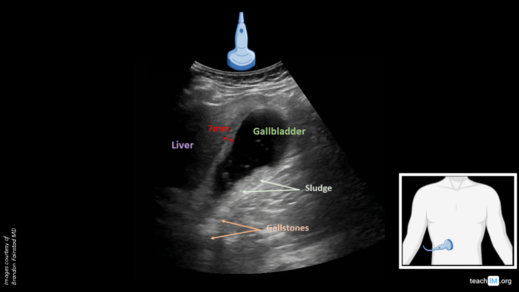
Cholecystitis – POCUS
Identify key biliary anatomy and differentiate between gallstones and biliary sludge based on an echogenic shaddow. (5 min)
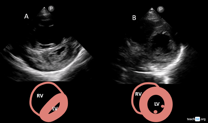
Right Ventricular (RV) Strain, “D-Sign” – POCUS
Identify key characterstics and generate a differential for right ventricular strain based on the parasternal short axis (PSAX) view. (5 min)
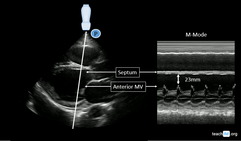
Reduced Systolic Function (EPSS) – POCUS
Identify systolic dysfuction in the PLAX view and use end-point septal seperation (EPSS) to characterize the severity (5 min case)
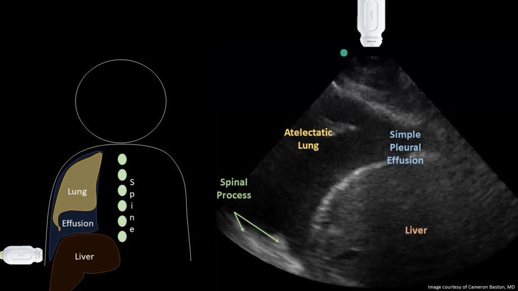
Pleural Effusion – POCUS
Identify a pleural effusion and using lung ultrasound and describe the relative diagnostic accuracy compared to chest radiograph. (5 minutes)
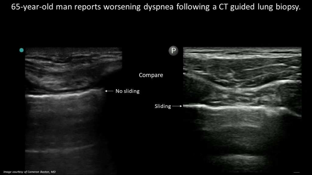
Pneumothorax (Lung Sliding) – POCUS
Identify the absence of lung sliding on ultrasound to diagnose a pneumothorax and review the anatomy of lung ultrasound (5 minutes)
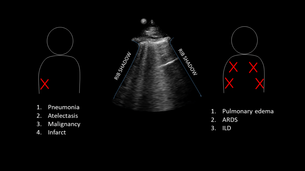
CHF Exacerbation (B-lines) – POCUS
Identify B-lines on lung ultrasound and generate a differential based on a clinical history and distribution (5 minutes).
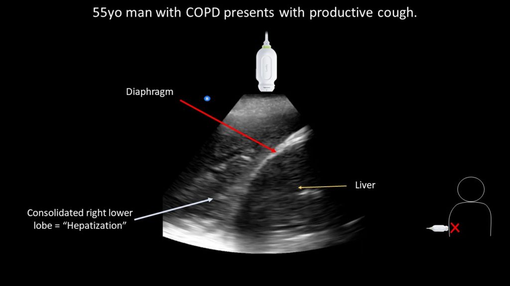
Pneumonia (“hepatization”) – POCUS
Use POCUS to identify a dense lobar consolidation ("hepatization") and distinguish the normal finding of "mirror artifact". (5 minutes).
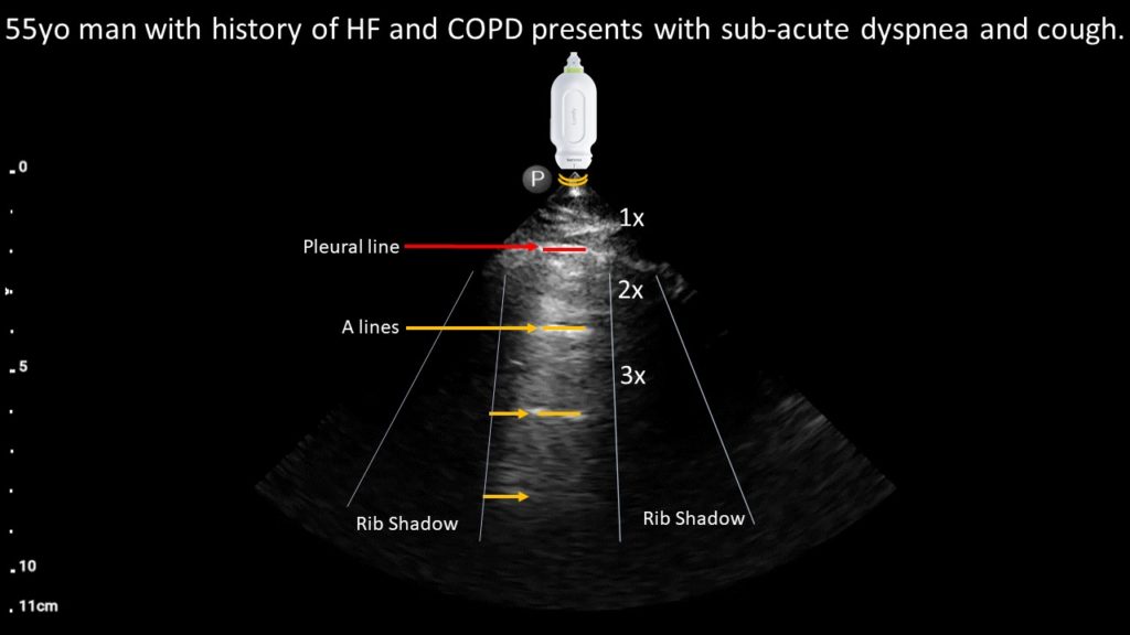
COPD Exacerbation (A-lines) – POCUS Case
Use POCUS to identify normal pleural and aerated lung in a patient with sub-acute dyspnea to diagnose a COPD exacerbation (5 minutes).
Case Conferences
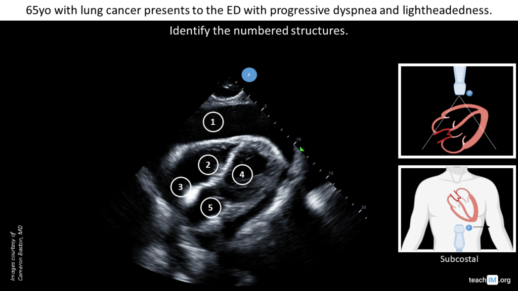
Cardiac Case Conference – POCUS
Introduce three standard cardiac ultrasound views through clinical integration of key cases: reduced EF, pericardial effusion, and RV strain.
Workshops
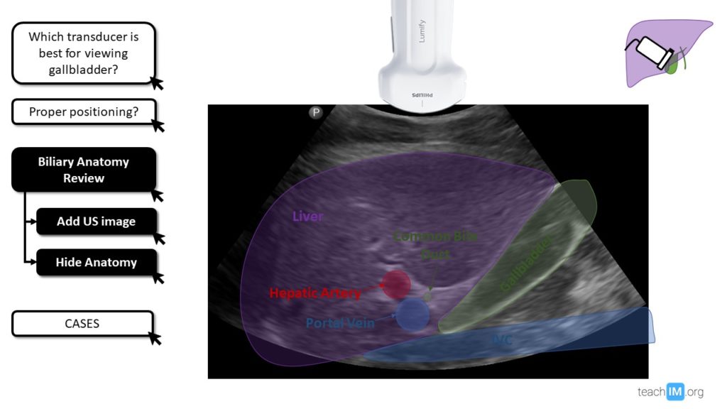
Galbladder Ultrasound
Practice best techniques for image acquisition, identify key anatomic structures and pathologic findings in gallbladder ultrasound. (15 min).
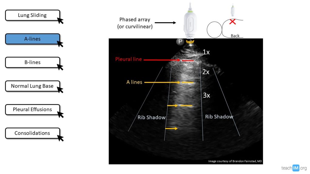
Lung and Pleural Ultrasound
Identify and describe lung sliding, A-lines, B-lines and lung consolidation using interactive graphics and videos (30 minutes)
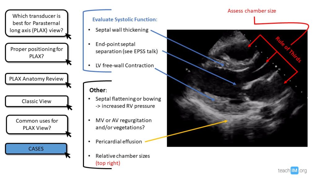
Parasternal Long Axis (PLAX) – POCUS
An interactive talk cases to demonstrate the basics of obtaining and interpreting a Parastenal Long Axis (PLAX or PSLA) view. (10-15 min)
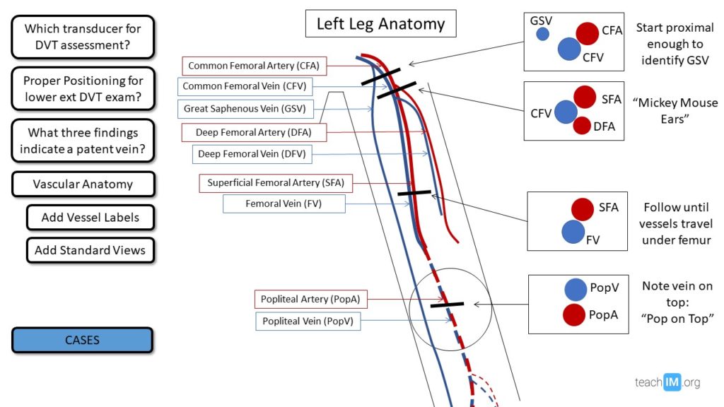
Deep Vein Thrombosis (DVT) – POCUS
Teach deep vein thrombosis (DVT) POCUS with pre-work videos, interactive presentation and instruction for hands-on training. (30min)