Published March 2021
Jennifer Doran, MD 1, Michelle Fleshner 2
1 Resident, Internal Medicine, University of Colorado, 2 Assistant Professor, Hospital Medicine, University of Colorado
Objective(s)
- Identify lung consolidation based on an echogenic pattern similar to the liver (“hepatization”) and differentiate this finding from the mirror artifact produced from viewing aerated lung through a hepatic window.
Teaching Instructions
Plan to spend 5-10 minutes familiarizing yourself with the animations of the PowerPoint and the key findings of this POCUS clip.
Instructions: Download the PowerPoint presentation file (videos do not work in the browser viewer) and have the image pulled up in presenter mode before learners look at the screen, to avoid revealing the diagnosis. Ask a learner to provide an overall interpretation. Then advance through the animations to prompt learners with key questions and reveal the findings, diagnosis, and teaching points. You can go back to prior graphics and questions by using the back arrow on the keyboard or by scrolling back on the mouse wheel.
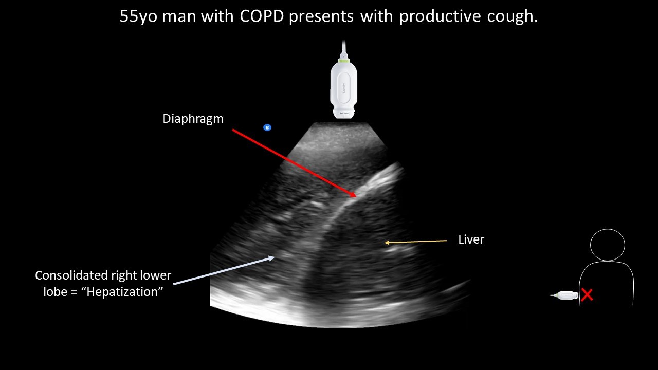
POCUS case image interpretation:
Right lower lobe consolidation or “hepatization” = dense lung tissue with visible airways that resemble biliary ducts of the liver.
Diagnosis: Likely community-acquired pneumonia.
Teaching:
- Aerated lung is not directly visualized by ultrasound due to the reflective properties of air.
- If the lung parenchyma is directly visualized this suggests increased density from atelectasis or consolidation.
- When the lung is densely consolidated the airways remain patent and well visualized, resembling the ultrasonographic appearance of the liver, referred to as “hepatization”.
- However, a similar appearance is seen in normal, well-aerated lungs, when the transducer footprint is placed directly over the liver. Using the liver as the echogenic window, ultrasound waves travel through the liver, diaphragm, and pleura before being reflected by air in the lung. This recurring reflection produces an artifact similar to A-lines, producing a recurring image of the liver deep to the pleura.
- “Mirror artifact” can be distinguished from “hepatization” by sliding the transducer superior to the diaphragm or by identifying discontinuation of visible vertebrae superior to the diaphragm.
Presentation Board
Take Home Point
- Lung parenchyma directly visualized by ultrasound is due to atelectasis or consolidation, the latter will resemble the appearance of a liver (“hepatization”)
- “Mirror artifact” is seen when the liver is used as the echogenic window and can be distinguished from “hepatization” by sliding the transducer superior to the diaphragm or by identifying discontinuation of visible vertebrae superior to the diaphragm.
References
Soni NJ. Point of care ultrasound. 2nd edition. ed. St. Louis, MO: Elsevier; 2019.