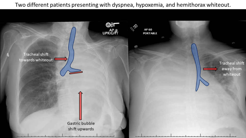
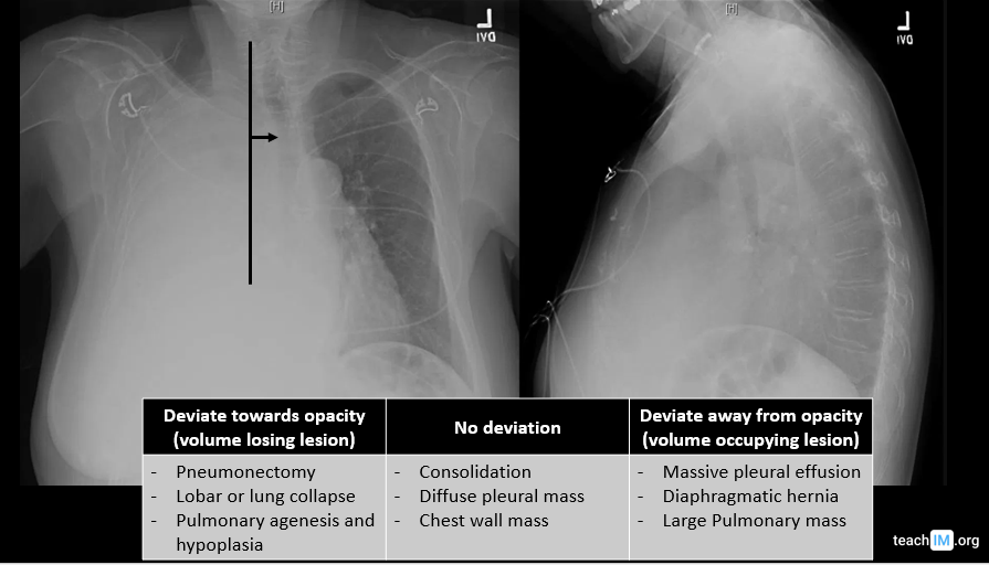
Hemithorax Whiteout – CXR Use tracheal deviation either toward or away from a large lung opacity on a CXR to narrow the differential of the lesion. (5 minutes)
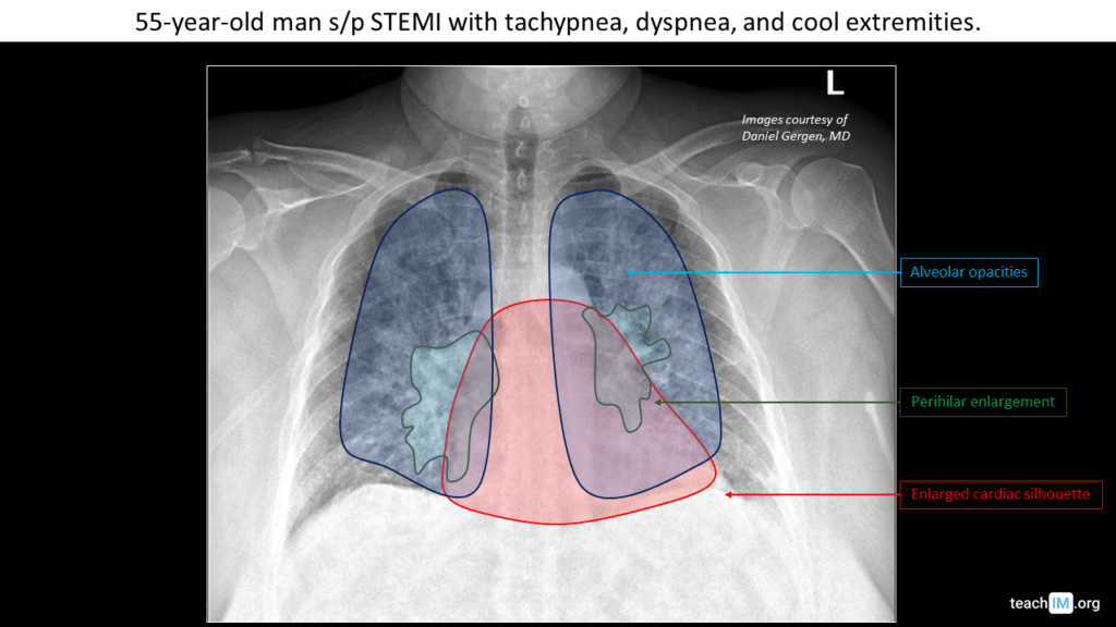
Left Heart Failure – CXR Identify key features to identify and determine the severity of congestive left heart failure on a chest x-ray. (5 minutes)
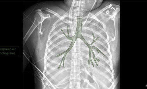
Air Bronchograms Learn to identify an air bronchogram on chest x-ray and describe the pathologies that lead to the appearance of air bronchograms.
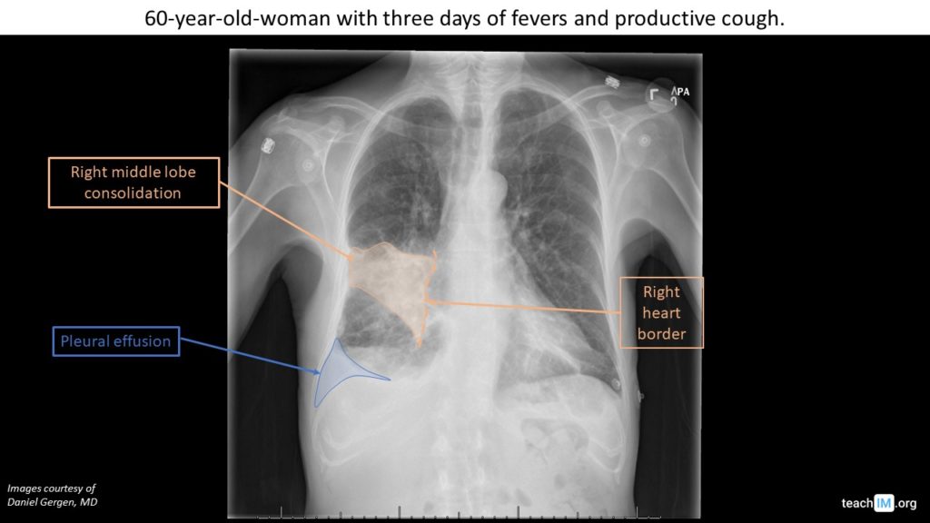
Silhouette Sign: RML Pneumonia – CXR Use the silhouette sign to localize a right middle lobe pneumonia and parapneumonic effusion on chest x-ray. (5 minutes)
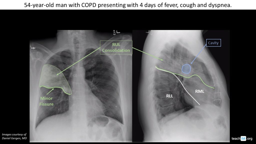
Bacterial Pneumonia with Abscess – CXR Use pleural fissures to determine the location of a right upper lobe pneumonia with a cavitary lung abscess. (5 minutes)
Malignant Pleural Effusion – CXR Use tracheal and mediastinal deviation to help differentiate the etiology of lung whiteout and other large opacities.
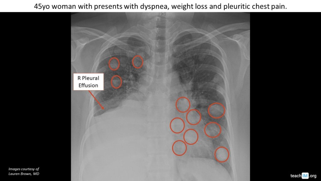
Lung Metastases, “Cannonball Lesions” – CXR Differentiate malignant metastases to the lung from primary lung malignancy. (5 min)
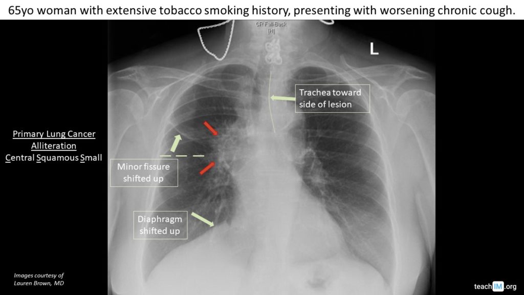
Hilar Mass – Primary Lung Cancer Identify key characteristics that differentiate a hilar mass due to primary lung cancer from other causes.
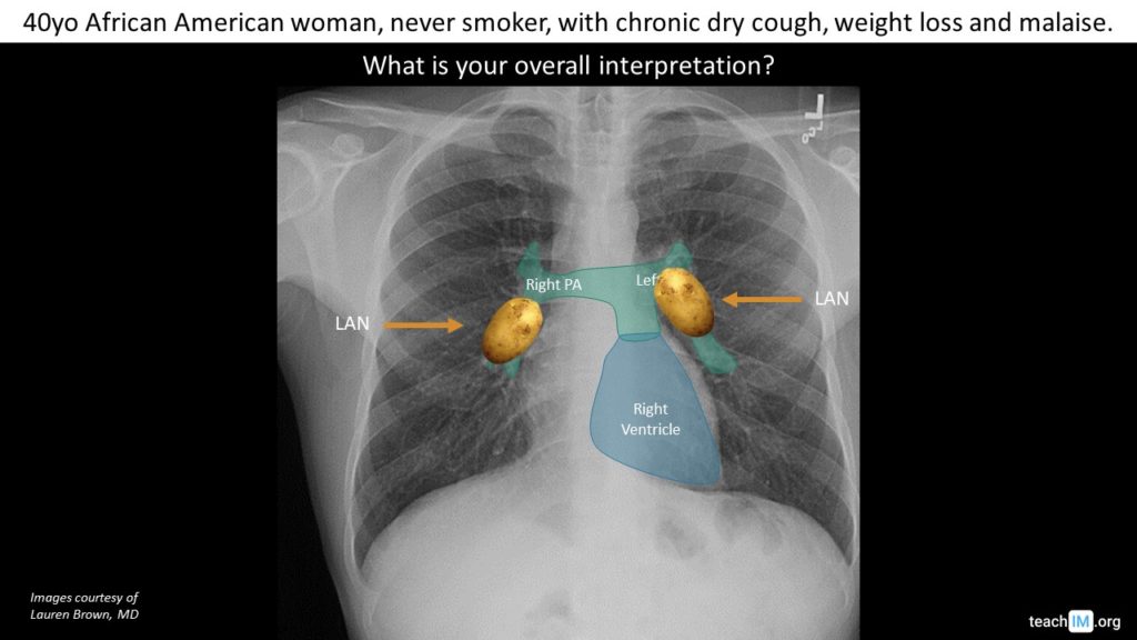
Hilar Lymphadenopathy Differentiate between bulky lymphadenopathy and enlarged pulmonary arteries in a CXR with enlarged hila.
Atelectasis – Left Lobar Collapse Identify mediastinal shift toward a lung opacification to diagnose atelectasis from lobar collapse.