Plan to spend 5-10 minutes familiarizing yourself with the animations of the PowerPoint and the key findings of this chest X-ray.
Instructions: Present the image either by expanding the window (bottom right) in a browser or downloading the PowerPoint file (downloading is recommended. Have the image pulled up in presenter mode before learners look at the screen to avoid revealing the diagnosis. Ask a learner to provide an overall interpretation. Then advance through the animations to prompt learners with key questions and reveal the findings, diagnosis, and teaching points. You can go back to prior graphics and questions by using the back arrow or scrolling back on the mouse wheel.
Official CXR Read: Dense right middle lobe consolidation and small right pleural effusion
Diagnosis: Right middle lobe pneumonia and parapneumonic effusion
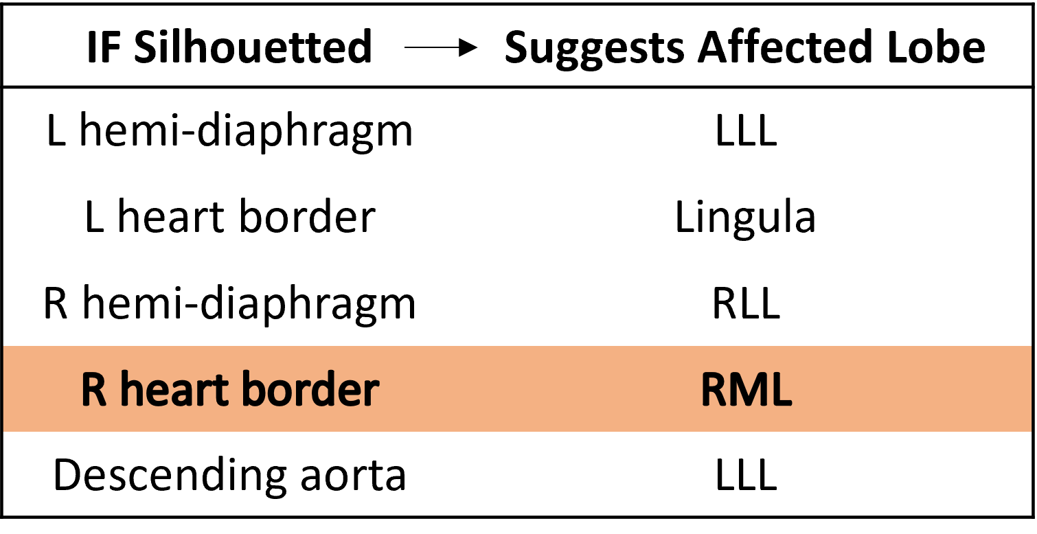
Teaching: A lung opacity in the mid-right lung fields with loss of the right heart border suggests a dense right middle lobe (RML) process. This is an example of the ‘silhouette sign’, the loss of a normal radiographic interface (e.g lung and mediastinum), due to similar density substances in direct contact. This makes it difficult to distinguish the borders of two tissues. A secondary finding is the small right pleural effusion. A pleural effusion adjacent to pneumonia is often due to inflammatory reactive fluid and termed a parapneumonic effusion. If the infection extends into the pleural effusion, it is classified as an empyema.