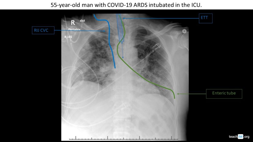
Gastric Tube Placement – CXR Use two rules to determine appropriate placement of a nasogastric or orogastric tube placement on a chest x-ray. (5 minutes)
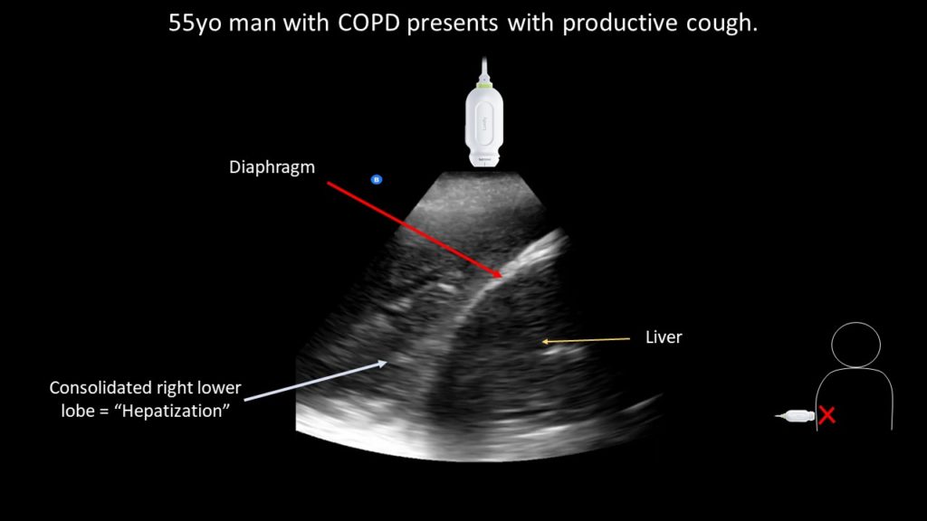
Pneumonia (“hepatization”) – POCUS Use POCUS to identify a dense lobar consolidation ("hepatization") and distinguish the normal finding of "mirror artifact". (5 minutes).
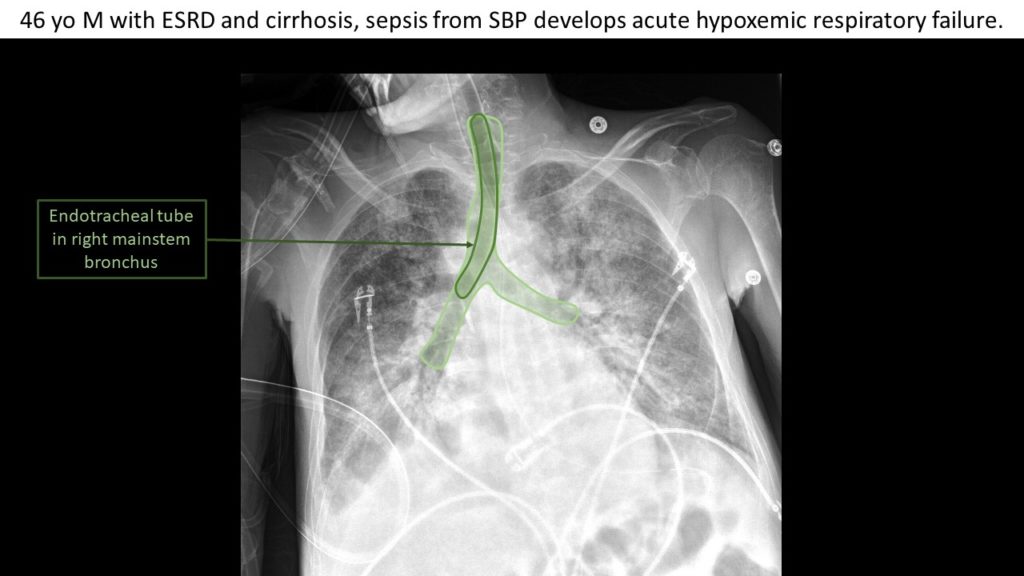
Right mainstem intubation – CXR Identify an endotracheal tube on chest x-ray and determine it's appropriate position.
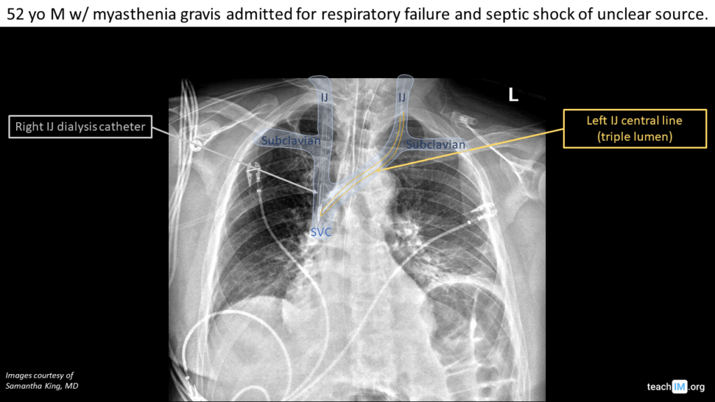
Central Line Placement – CXR Identify left and right central venous catheters (CVCs) on a chest x ray and the appropriate location of their placement
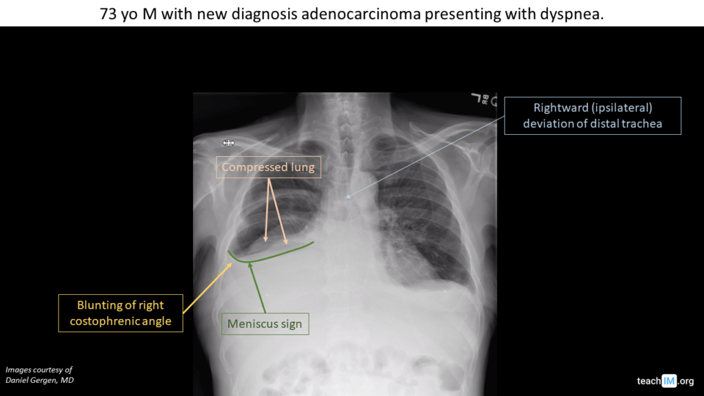
Pleural Effusion – CXR Use the meniscus sign to identify a pleural effusion. Use the degree of mediastinal shift to determine preponderance of effusion vs. atelectasis.
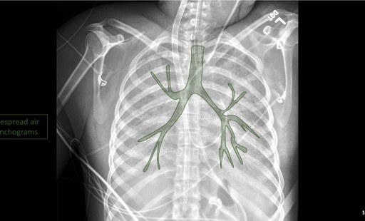
Air Bronchograms Learn to identify an air bronchogram on chest x-ray and describe the pathologies that lead to the appearance of air bronchograms.
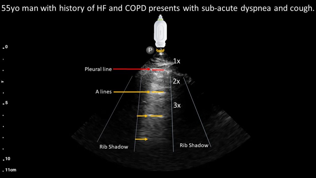
COPD Exacerbation (A-lines) – POCUS Case Use POCUS to identify normal pleural and aerated lung in a patient with sub-acute dyspnea to diagnose a COPD exacerbation (5 minutes).
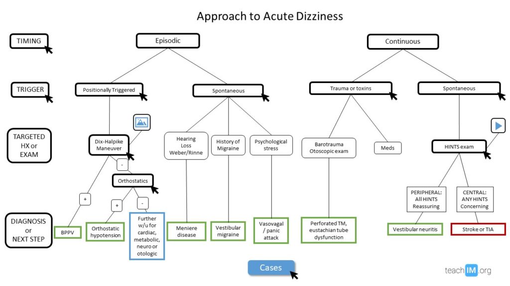
Acute Dizziness Use a four-step bedside approach to differentiate between benign and central causes of dizziness. (45 minutes)
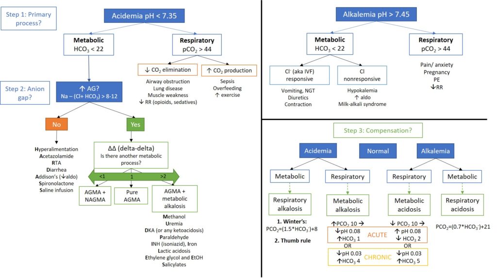
Acid Base Disorders Develop an approach to identifying acid-base disorders and practice the interpretation of ABGs through a series of cases (45 minutes).
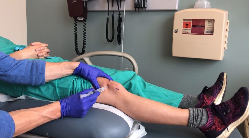
Knee Arthrocentesis Comprehensive procedural training for knee injection and aspiration through both simulation and clinical instruction. (45 min)