Published July 2021
Brandon Fainstad, MD
Expert review – pending
Assistant Professor, General Internal Medicine, University of Colorado
Objective(s)
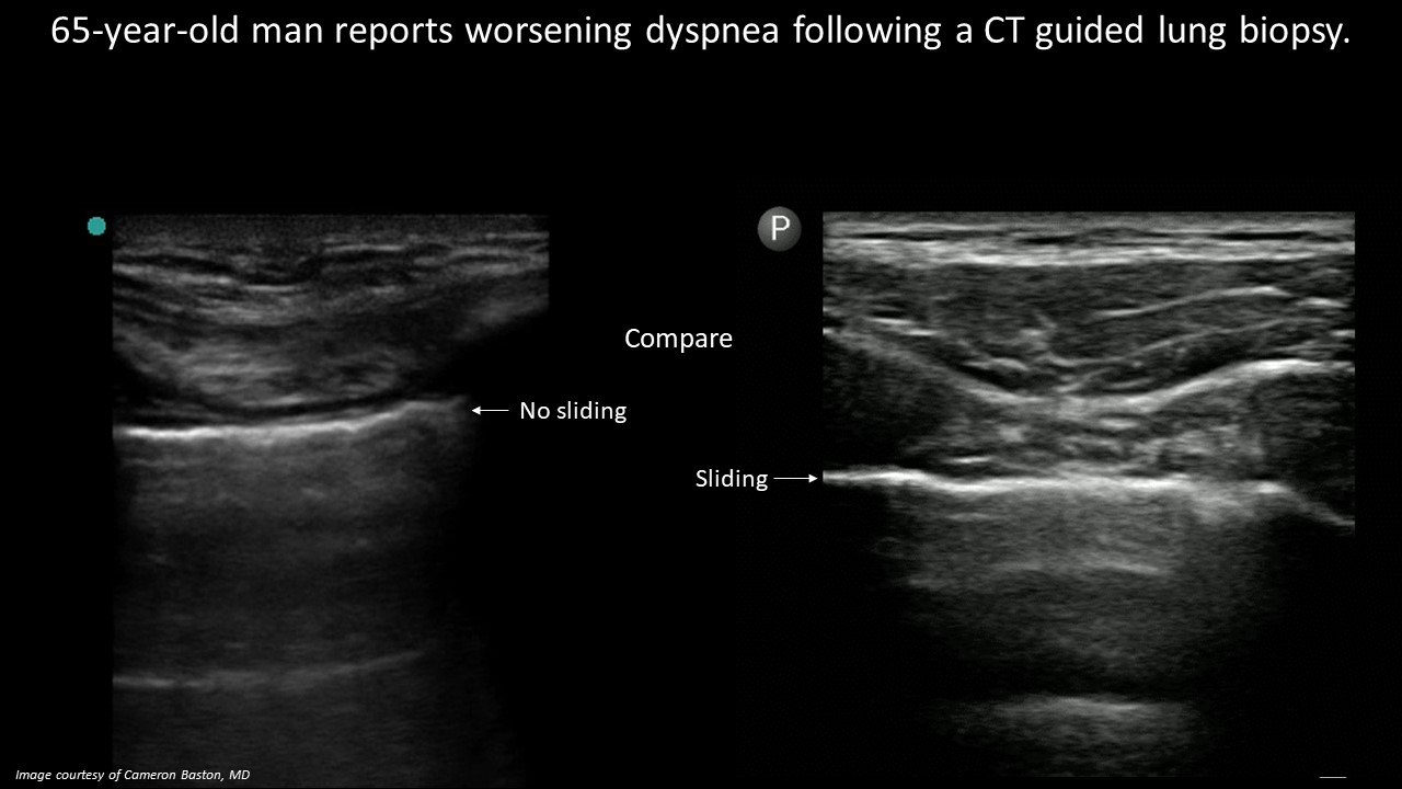
- Identify the absence of lung sliding on lung ultrasound to narrow the differential for dyspnea.
- Describe the anatomy visualized on lung ultrasound.
Teaching Instructions
Plan to spend 5-10 minutes familiarizing yourself with the animations of the PowerPoint and the key findings of this POCUS clip.
Instructions: Download the PowerPoint presentation file (videos do not work in the browser viewer) and have the image pulled up in presenter mode before learners look at the screen, to avoid revealing the diagnosis. Ask learners to discuss with their neighbors the answer to each question for about 30 seconds. Then advance through the animations and teaching points to the next question, and repeat. You can go back to prior graphics and questions by using the back arrow on the keyboard or by scrolling back on the mouse wheel. This should take no more than 5-10 mintues.
Diagnosis: Pneumothorax.
Ultrasound interpretation: Static lung pleura with active intercostal movements.
Teaching:
A healthy and functional interface of the visceral and parietal lung pleura slide against one another during respiration. This is observed by ultrasound with a sliding or ‘shimmering’ (or ‘ants on a long’) at the level of the pleura and deep to it. It is seen as the ‘seashore sign’ on the one-dimensional M-mode. If any portion of this process is interrupted (no lung movement, air between the two layers, or adhesions between the two layers) there is a notable absence of lung sliding.
Presentation Board
Take Home Point
- Sliding or ‘shimmering’ of the lung pleura suggests the two layers are sliding against one another and rules out a pneumothorax for that specific location.
- The absence of lung sliding suggests any interruption in the process of lung sliding (no lung movement, air between the layers or adhesions between the layers).
References
Soni NJ. Point of care ultrasound. 2nd edition. ed. St. Louis, MO: Elsevier; 2019