Chalk Talks
Cardiology
Endocrinology
Gastroenterology
Hematology
Immunology
Infectious Disease
Nephrology
Neurology
Oncology
Primary Care
Pulmonology
Rheumatology
Transplant Medicine
Women’s Health
Procedures
Curricula Overviews
Procedures
POCUS
5-minute Cases
Case Conferences
Workshops
CXR
Case Conferences
Cavitary Lesion
Interstitial Lung Disease
Mediastinum and Hila
Obstructive lung diseases
Opacities
Pleural
Tubes, Lines, and Drains
ECG
Atrioventricular (AV) Heart Blocks
Bradycardias
Bundle Branch Block
Case Conferences
Electrolyte Abnormalities
Introduction
ST changes
Supraventricular Tachycardia
Dermatology
About
About Us
Contact
Twitter
Chalk Talks
Cardiology
Endocrinology
Gastroenterology
Hematology
Immunology
Infectious Disease
Nephrology
Neurology
Oncology
Primary Care
Pulmonology
Rheumatology
Transplant Medicine
Women’s Health
Chest x-ray
Case Conferences
Cavitary Lesion
Interstitial Lung Disease
Mediastinum and Hila
Obstructive lung diseases
Opacities
Pleural
Tubes, Lines, and Drains
ECGs
Atrioventricular (AV) Heart Blocks
Bradycardias
Bundle Branch Block
Case Conferences
Electrolyte Abnormalities
Introduction
ST changes
Supraventricular Tachycardia
POCUS
5-minute Cases
Case Conferences
Workshops
Procedures
Curricula Overviews
Procedures
About Us
Contact
Chest x-ray
Case Conferences
Central Chest Opacities – CXR
Differentiate between various central chest opacities based on silhouettes and overlay of mediastinal structures (45 min)
Discrete Lung Opacites – CXR
Diffentiate varoius discrete lung opacities, including cavitary lesions, lung metastases, pulmonary nodules, lobar pneumnias and retrocardiac opacities (6 cases, 45 minutes)
Large and Diffuse Opacities – CXR
Differentiate large lung opacities based on the distribution, shift of adjacent structures, and silhouette signs. (5 cases, 40 min)
Lines, Fissures and Silhouettes – CXR
Introduce key concepts of fissures, silhouettes and invasive tubes, lines and devices on CXRs with case-based conference (45 min)
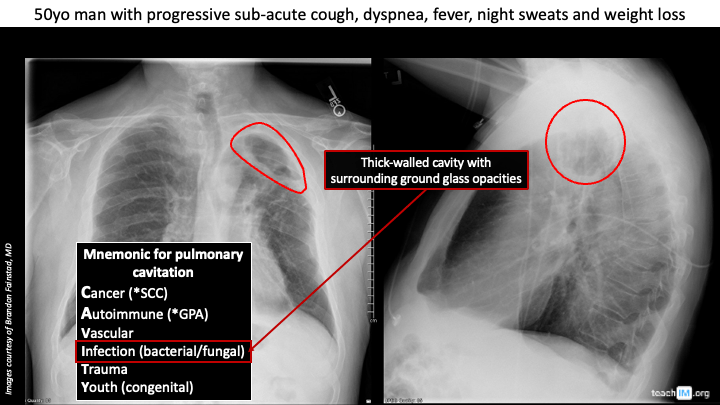
Cavitary Lesion
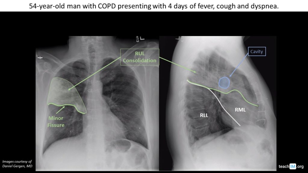
Bacterial Pneumonia with Abscess – CXR
Use pleural fissures to determine the location of a right upper lobe pneumonia with a cavitary lung abscess. (5 minutes)
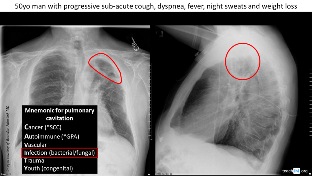
Actinomyces Lung Abscess – CXR
Identify distinguishing features and useful mnemonic for cavitary lung lesions.
Interstitial Lung Disease
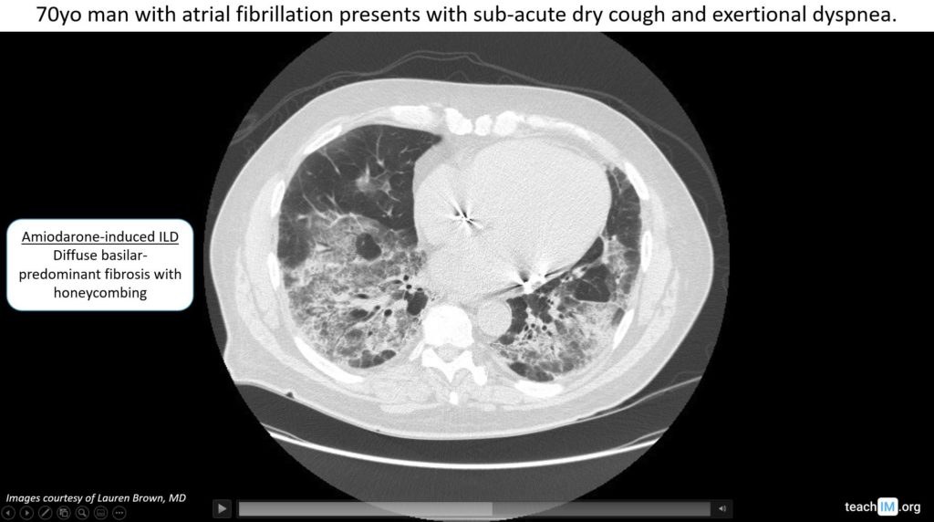
Amiodarone-Induced ILD
Identify reticular opacities on a CXR due to amiodarone-induced interstitial lung disease.
Mediastinum and Hila
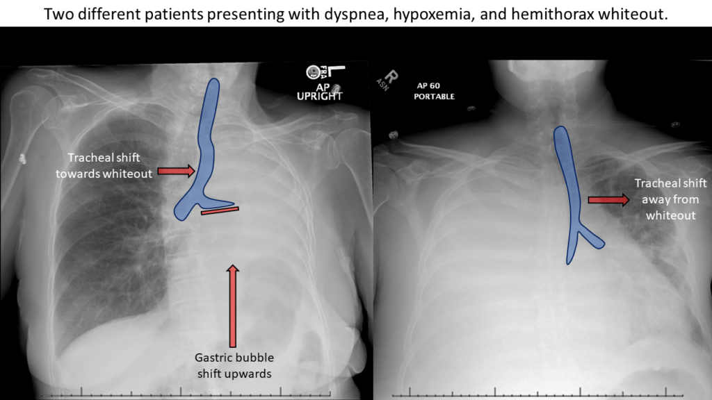
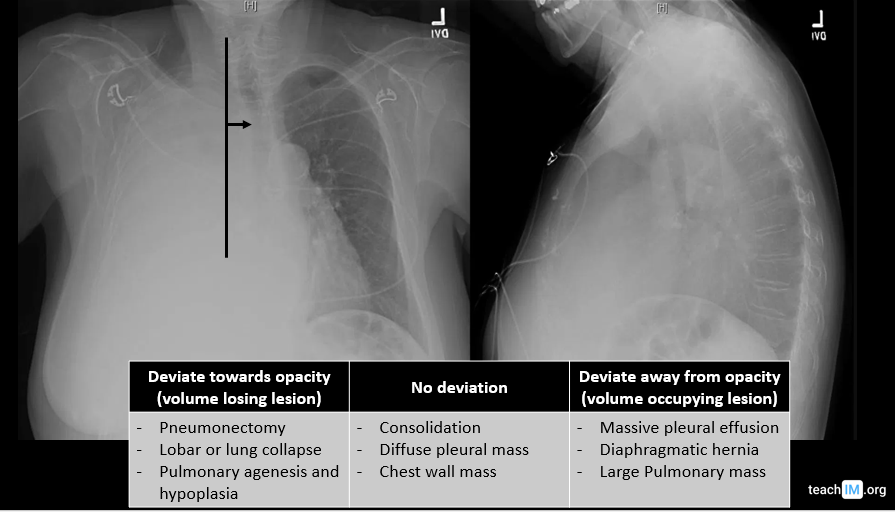
Hemithorax Whiteout – CXR
Use tracheal deviation either toward or away from a large lung opacity on a CXR to narrow the differential of the lesion. (5 minutes)
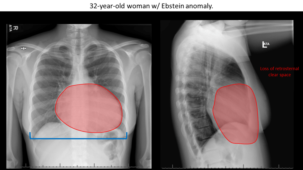
Cardiomegaly (Right Heart) – CXR
Identify an enlarged cardiac silhouette and determine the cardiac chambers accounting for cardiomegaly on a CXR. (5 minutes)
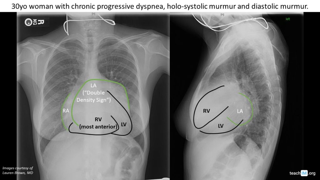
Left Atrial Enlargement
Identify cardiomegaly with enlarged cardiac silhouette predominantly due to left and right atrial enlargement. (5 minutes)
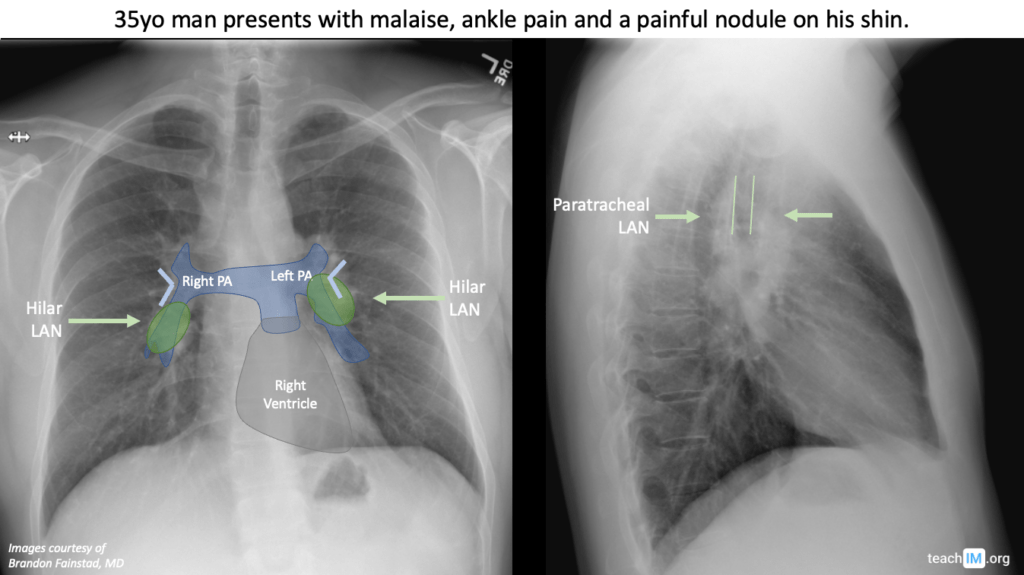
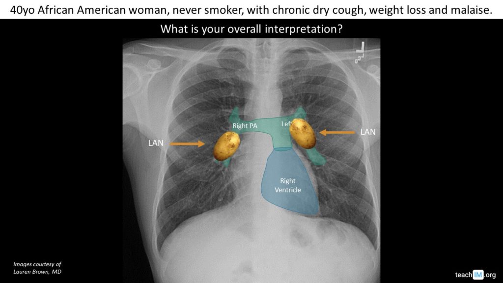
Hilar Lymphadenopathy
Differentiate between bulky lymphadenopathy and enlarged pulmonary arteries in a CXR with enlarged hila.
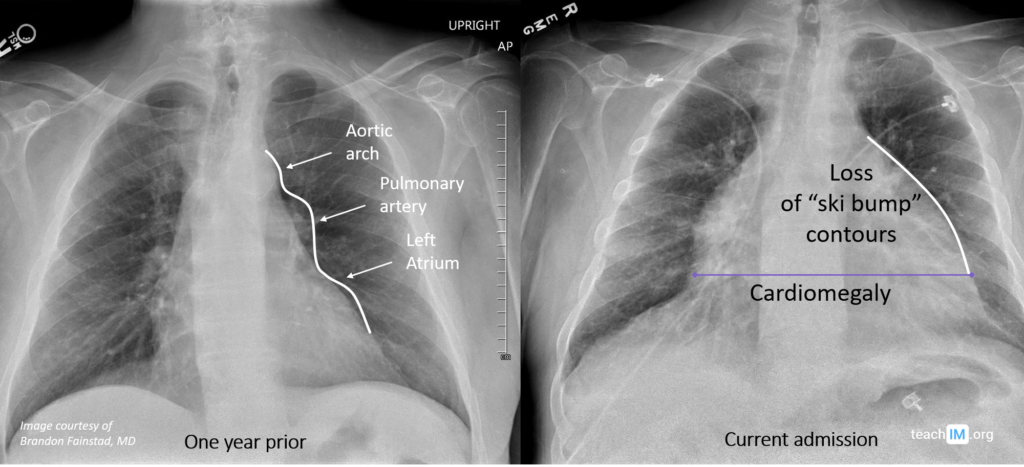
Pericardial Effusion
Identify key features of a cardiac silhouette suggestive of a pericardial effusion
Obstructive lung diseases
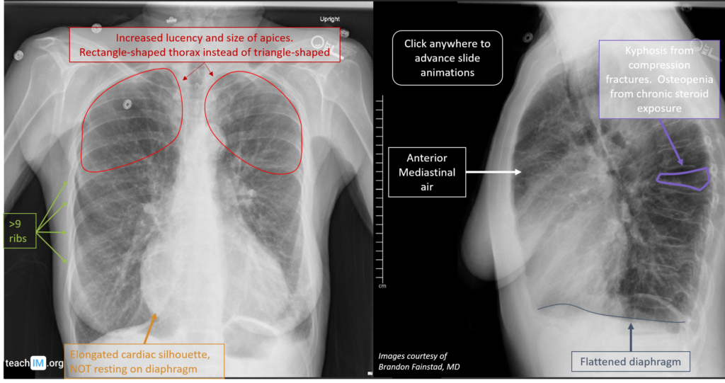
Excipient Lung Disease (Basilar Emphysema) – CXR
Differentiate basilar from apical emphysema to demonstrate the pathophysiologic difference from hematogenous versus inhaled lung toxicity.
Chronic Obstructive Pulmonary Disease (COPD)
Identify key radiographic features to COPD and apical emphysema.
Opacities
Hemithorax Whiteout – CXR
Use tracheal deviation either toward or away from a large lung opacity on a CXR to narrow the differential of the lesion. (5 minutes)
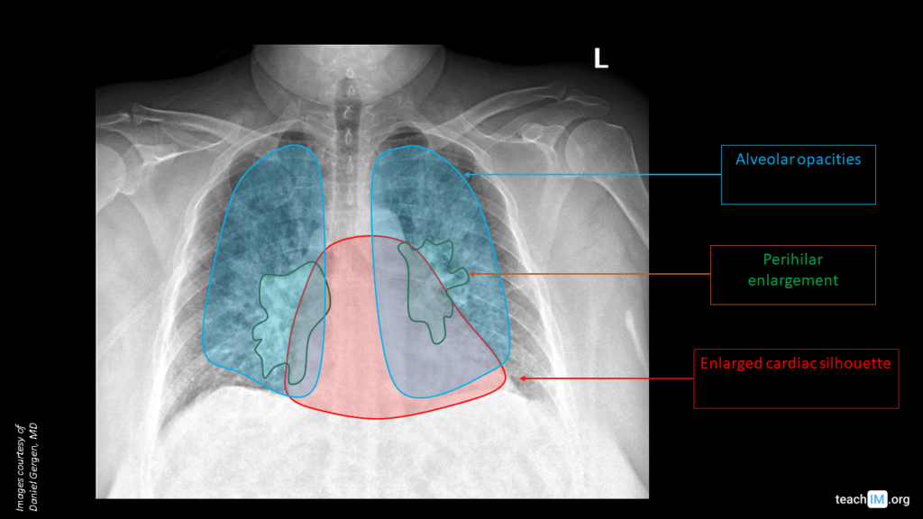
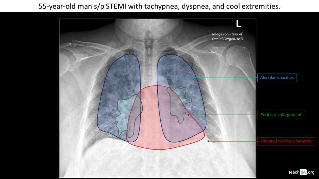
Left Heart Failure – CXR
Identify key features to identify and determine the severity of congestive left heart failure on a chest x-ray. (5 minutes)
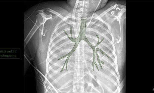
Air Bronchograms
Learn to identify an air bronchogram on chest x-ray and describe the pathologies that lead to the appearance of air bronchograms.
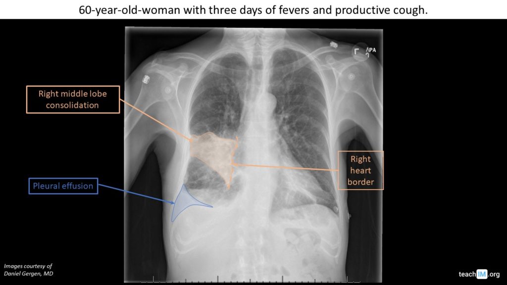
Silhouette Sign: RML Pneumonia – CXR
Use the silhouette sign to localize a right middle lobe pneumonia and parapneumonic effusion on chest x-ray. (5 minutes)
Bacterial Pneumonia with Abscess – CXR
Use pleural fissures to determine the location of a right upper lobe pneumonia with a cavitary lung abscess. (5 minutes)
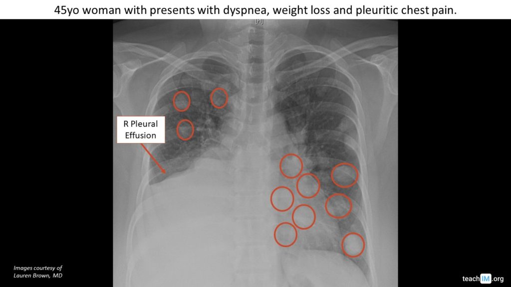
Malignant Pleural Effusion – CXR
Use tracheal and mediastinal deviation to help differentiate the etiology of lung whiteout and other large opacities.
Lung Metastases, “Cannonball Lesions” – CXR
Differentiate malignant metastases to the lung from primary lung malignancy. (5 min)
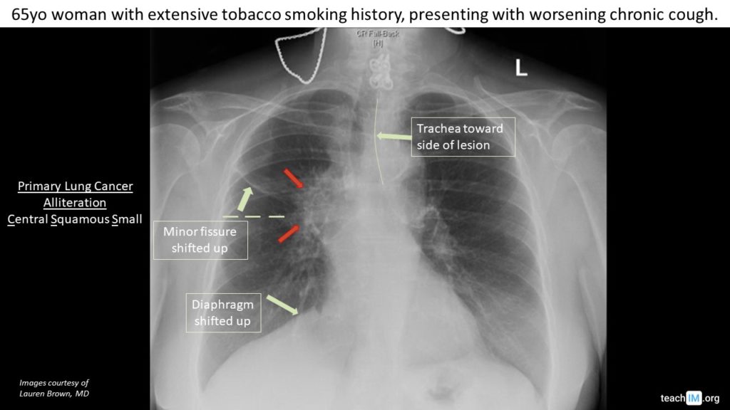
Hilar Mass – Primary Lung Cancer
Identify key characteristics that differentiate a hilar mass due to primary lung cancer from other causes.
Hilar Lymphadenopathy
Differentiate between bulky lymphadenopathy and enlarged pulmonary arteries in a CXR with enlarged hila.
Atelectasis – Left Lobar Collapse
Identify mediastinal shift toward a lung opacification to diagnose atelectasis from lobar collapse.
Actinomyces Lung Abscess – CXR
Identify distinguishing features and useful mnemonic for cavitary lung lesions.
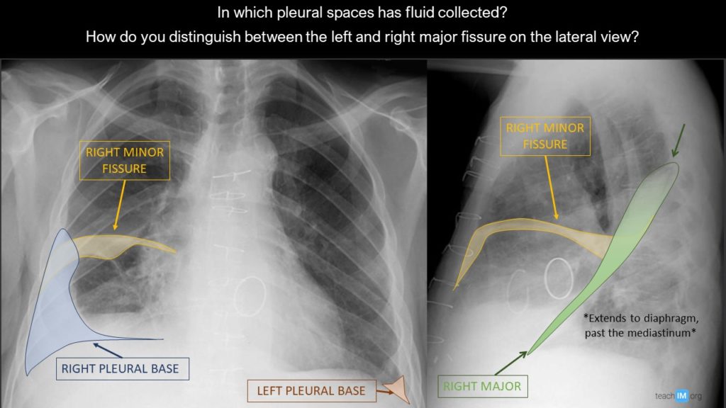
Pleural
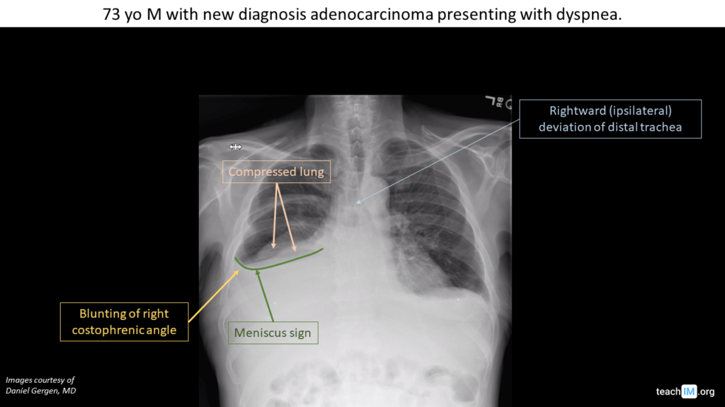
Pleural Effusion – CXR
Use the meniscus sign to identify a pleural effusion. Use the degree of mediastinal shift to determine preponderance of effusion vs. atelectasis.
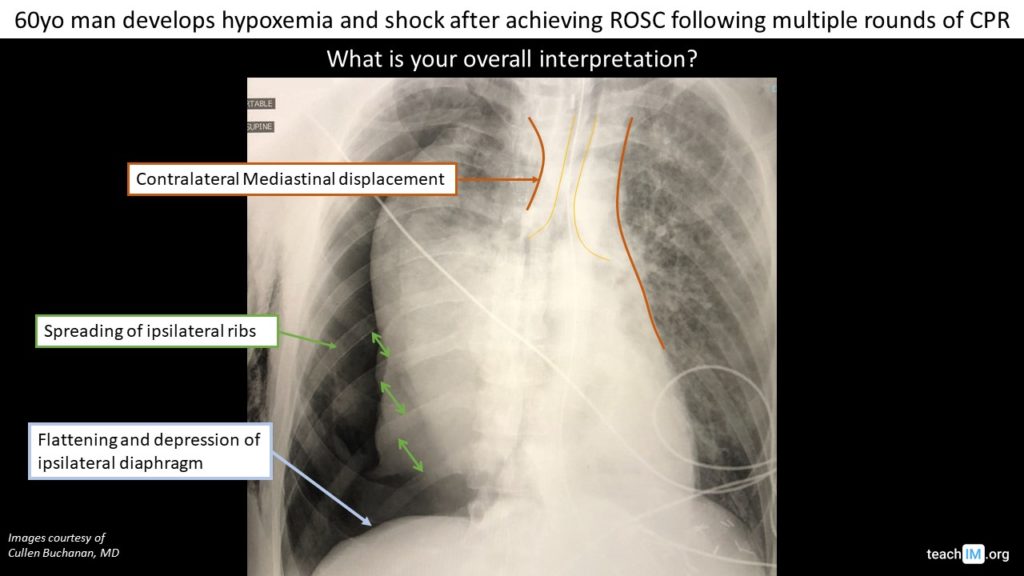
Tension Pneumothorax – CXR
Identify key features on a CXR that distinguish a tension pneumothorax from a simple pneumothorax on a (5 minutes)
Malignant Pleural Effusion – CXR
Use tracheal and mediastinal deviation to help differentiate the etiology of lung whiteout and other large opacities.
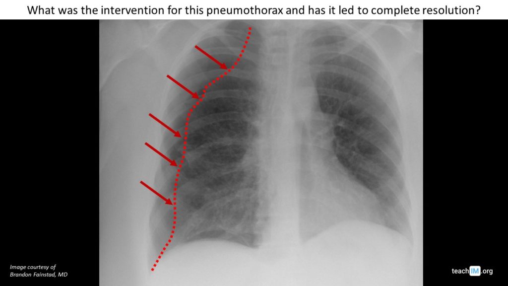
Pneumothorax
Identify a pneumothorax and assess response to decompression with a chest tube.
Lobes and Fissures
Identify pleural effusions in multiple fissures
Tubes, Lines, and Drains
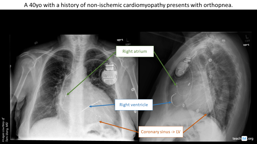
Pacemakers and Defibrillators – CXR
Determine the anatomic location of intracardiac leads and differentiate between a pacemaker and an ICD based on CXR. (5 min)
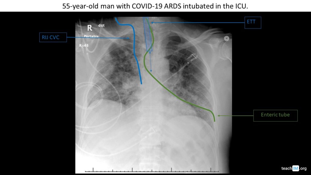
Gastric Tube Placement – CXR
Use two rules to determine appropriate placement of a nasogastric or orogastric tube placement on a chest x-ray. (5 minutes)
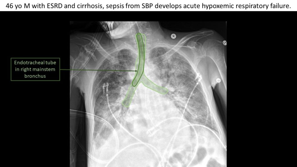
Right mainstem intubation – CXR
Identify an endotracheal tube on chest x-ray and determine it's appropriate position.
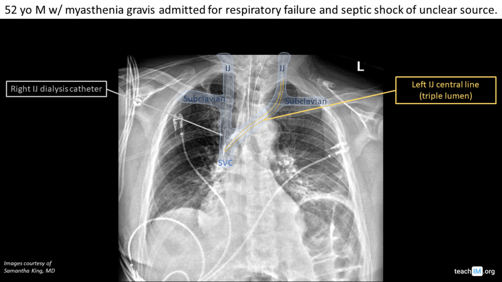
Central Line Placement – CXR
Identify left and right central venous catheters (CVCs) on a chest x ray and the appropriate location of their placement