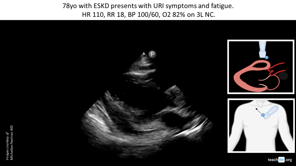
Pericardial Effusion Without Tamponade – POCUS Identify a pericardial effusion in the parasternal long (PLAX) and assess for clinical signs of tamponade. (5 minutes)
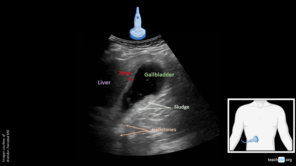
Cholecystitis – POCUS Identify key biliary anatomy and differentiate between gallstones and biliary sludge based on an echogenic shaddow. (5 min)
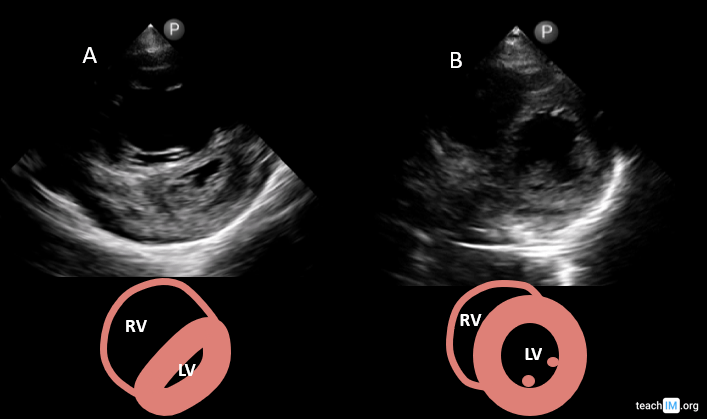
Right Ventricular (RV) Strain, “D-Sign” – POCUS Identify key characterstics and generate a differential for right ventricular strain based on the parasternal short axis (PSAX) view. (5 min)
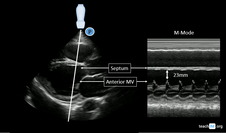
Reduced Systolic Function (EPSS) – POCUS Identify systolic dysfuction in the PLAX view and use end-point septal seperation (EPSS) to characterize the severity (5 min case)
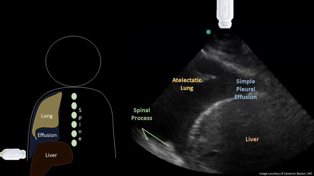
Pleural Effusion – POCUS Identify a pleural effusion and using lung ultrasound and describe the relative diagnostic accuracy compared to chest radiograph. (5 minutes)
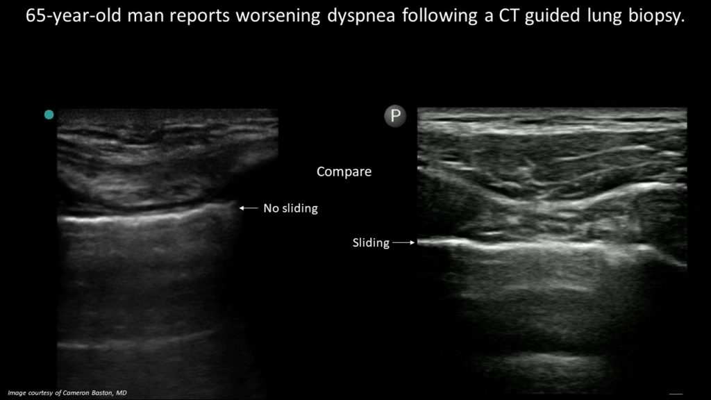
Pneumothorax (Lung Sliding) – POCUS Identify the absence of lung sliding on ultrasound to diagnose a pneumothorax and review the anatomy of lung ultrasound (5 minutes)
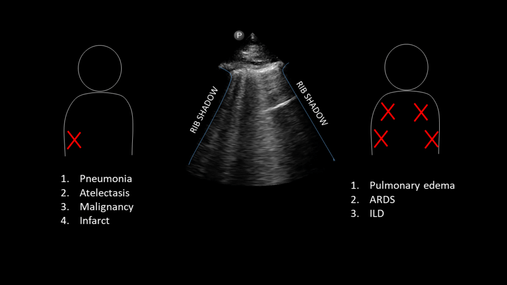
CHF Exacerbation (B-lines) – POCUS Identify B-lines on lung ultrasound and generate a differential based on a clinical history and distribution (5 minutes).
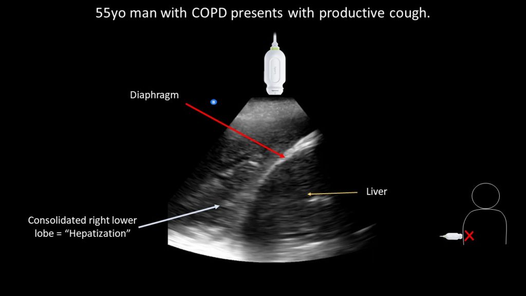
Pneumonia (“hepatization”) – POCUS Use POCUS to identify a dense lobar consolidation ("hepatization") and distinguish the normal finding of "mirror artifact". (5 minutes).
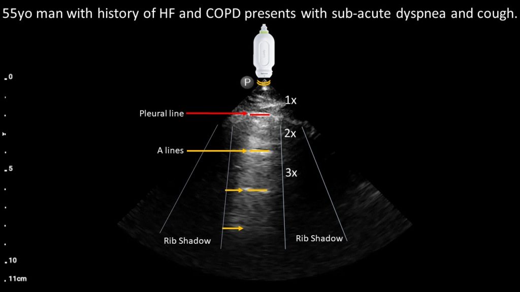
COPD Exacerbation (A-lines) – POCUS Case Use POCUS to identify normal pleural and aerated lung in a patient with sub-acute dyspnea to diagnose a COPD exacerbation (5 minutes).